
Pet Dental X-Ray: Why Your Vet Wants One Before Cleaning
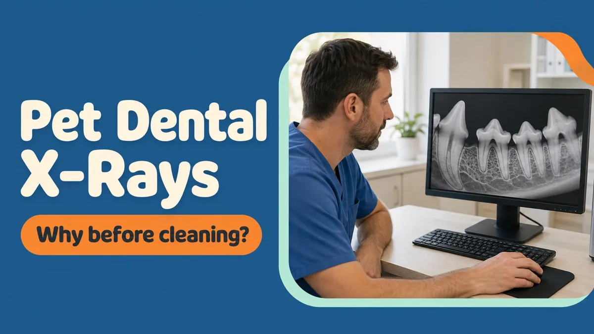
Owners are reasonably suspicious of anything added to a quote, and a pet dental x-ray before a cleaning sounds exactly like one of those additions. It is a fair instinct, and in this case it points the wrong way — the images are not an extra layer of thoroughness on top of the procedure. They are the part that decides what the procedure is.
What follows explains what the pictures show, why they are taken while he is already asleep, and why an estimate given before them is a rougher number than most clinics admit.
Two-thirds of a tooth is somewhere you cannot look
Roughly two-thirds of every tooth sits below the gumline: the root, the ligament that suspends it, and the bone that holds the whole arrangement in place. Everything visible in the mouth — the part you would see if you lifted a lip — is the remaining third.
Almost all serious dental disease happens in the hidden two-thirds. Infection at the tip of a root, bone dissolving around a tooth that still looks solid, a root fractured under an intact crown, a tooth being resorbed from within. None of it changes the appearance of the crown until very late, which is why a mouth can be graded as mild from the outside and turn out to be nothing of the kind.
What the pictures change
In practice, the radiographs alter the plan more often than any other step in the day. They do it in both directions, and the second one is the part owners do not expect.
They add work when a tooth that looked acceptable turns out to have lost the bone around its root — that tooth is not saved by scaling, it is a source of infection that has to come out. And they remove work just as often: a tooth that looks alarming, heavily stained and surrounded by inflamed gum, can be perfectly sound underneath, in which case it is cleaned and kept rather than extracted. Without the images, the safe assumption tends to be extraction, and teeth are removed that did not need to be.
They also explain the behaviour you noticed at home. Whatever it was, there is usually a specific tooth behind it, and the radiograph is where that tooth is finally named. The full picture of how the mouth is assessed from the beginning is on our dental service page.
Why it happens under anaesthesia
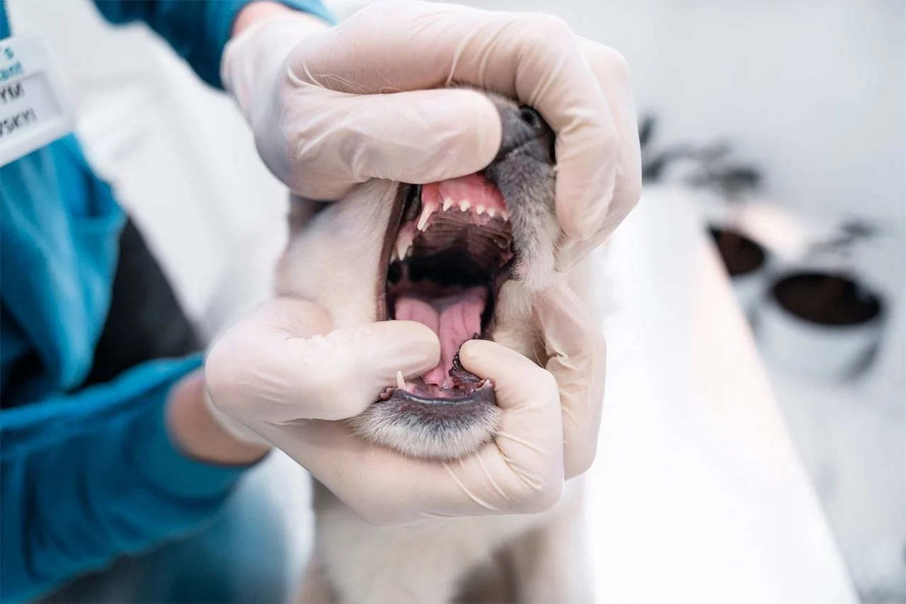
A dental radiograph requires a small sensor to be held inside the mouth, in a precise position, while the animal stays completely still. No conscious dog or cat tolerates this, and the alternative — approximate images taken from outside the mouth — will not show what is being looked for.
This is also the honest answer to the anaesthesia-free cleaning offered around the city. Scraping visible tartar off a crown while an animal is held still produces a whiter tooth and leaves the disease entirely untouched, because the disease is below the gumline where no awake animal will allow an instrument to go. It looks like treatment and it treats nothing.
Since he has to be asleep for proper scaling below the gumline in any case, the radiographs cost him nothing extra in anaesthetic time worth mentioning. Taking them in a second session would mean a second anaesthetic, which is the outcome everyone wants to avoid.
Why the number can move once the images are up
Clinics in this city are asked for a fixed price on the phone constantly, and the pressure to give one is real. We give a published price for the cleaning itself and a written estimate for the rest — and we say plainly that the estimate for extractions and gum treatment is provisional until the x-rays are on the screen.
That is not a hedge, it is the sequence. The mouth is examined, the blood work is run, he is anaesthetised, the images are taken, and only then does anyone know how many teeth are involved. What we do at that point is call you with the finding and the revised number before the work begins, so the decision stays yours rather than being reported afterwards.
It happens in this building
The radiographs are taken and read here in Al Quoz 3, on our own digital equipment, during the same admission. Nothing is sent out, nobody is referred to a second centre mid-procedure, and there is no second appointment to arrange in a city where crossing town for an imaging slot can cost a working day. The wider imaging service, including the chest and abdomen work that has nothing to do with teeth, is on our digital x-ray page.
What it costs
Full-mouth radiographs are part of a dental at Petbook rather than a line added to the bill — there is no separate imaging charge for the mouth, and no second invoice arriving later from somewhere else. What a dental itself costs for a cat and for a dog, and what the consultation before it costs, are set out on our published price list. The one figure that cannot be printed in advance is the extraction and gum work, for the reason above.
Ask to see them
You are entitled to look at the images and to have them explained, and a tooth that was extracted should be visible on a radiograph as a tooth that needed to come out. We go through them at discharge, which is generally the moment a dental stops feeling like an expense and starts making sense — most owners have never seen the inside of their animal’s jaw, and the difference between a healthy root and a diseased one is obvious to anybody once it is pointed out.
It is also the point at which the home-care conversation becomes worth having, because you now know which teeth are left and which of them are under pressure. What a full dental day looks like for a cat, from admission to discharge, is on our cat dental cleaning page.